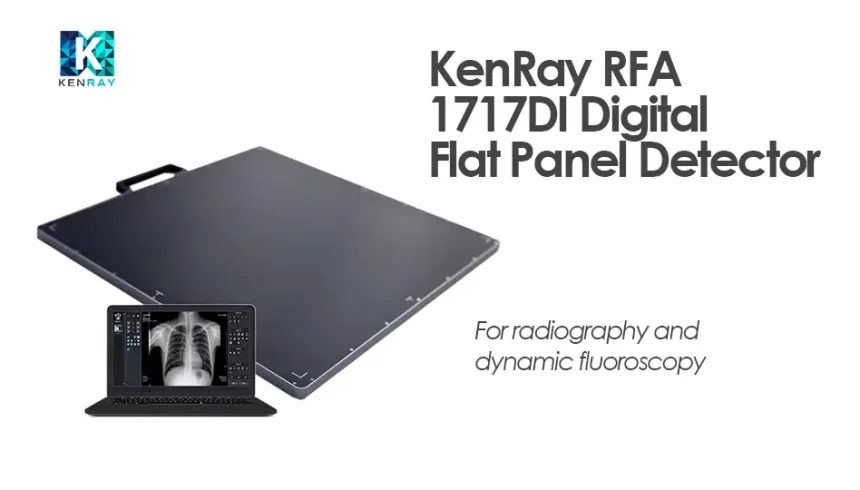
In an era where digital imaging is rapidly replacing traditional film radiography, the KenRay RFA‑1717DI stands out as a sophisticated, high-performance flat panel detector (FPD) that elevates both diagnostic quality and operational efficiency. Supporting both static X-ray imaging and real-time fluoroscopy, this 17 × 17″ cassette-sized detector is designed for radiology departments upgrading their analog systems or expanding into dynamic imaging workflows.
Product Overview
The KenRay RFA‑1717DI (also known as RFA-1717DIG for Gd₂O₂S or RFA-1717DIC for CsI scintillator) offers an active imaging area that adheres to ISO 4090 cassette standards (approximately 430×430 mm), enabling seamless retrofit into existing X-ray rooms without altering bucky trays or room layout.
With a high-resolution 3072×3072 pixel matrix and 140 µm pixel pitch, this detector achieves a spatial resolution of approximately 3.4 lp/mm, ideal for revealing fine anatomical details even under low-dose exposure conditions.
Uniquely, the RFA‑1717DI supports dynamic fluoroscopy up to 30 frames per second (at 1536×1536 resolution, 2×2 binning), making it versatile for procedures ranging from gastrointestinal motility studies to interventional guidance.
Core Technical Features
Imaging Performance
- Pixel Matrix: 3072 × 3072
- Spatial Resolution: ~3.4 lp/mm (16-bit grayscale, 65,536 gray levels)
- Scintillator Options: Gd₂O₂S:Tb (Gadox) or CsI:Tl for variable sensitivity and dose optimization.
Fluoroscopy & Frame Rates
Maximum Frame Rates:
- 30 fps @ 1536×1536 (2×2)
- 36 fps @ 1536×1024
- 50 fps @ 768×768
- Up to 84 fps in tighter ROIs (e.g., 768×128).
Auto Exposure Detection (AED)
- Integrated AED and line trigger support, enabling synchronization with X-ray generators for efficient static and pulsed acquisition without manual triggering setups.
Physical and Connectivity Specs
- Digital Sensor Size: 430.08 × 430.08 mm
- Weight: ~3.4 kg (Gadox) or ~3.5 kg (CsI), with a thickness of ~15 mm for cassette compatibility.
- Communications: Gigabit Ethernet (1000Base‑T); TTL sync ports for READY/EXPOSURE control.
- Power: DC 15 V via RFA‑PCON control unit
Designed for Clinical Versatility
Retrofit Radiography Upgrades
The RFA‑1717DI transforms analog radiography suites into digital imaging rooms, maintaining bucky tray compatibility with minimal installation changes.
Portable Fluoroscopy & Interventional Use
With its slim design and AED sync support, the detector is suited for fixed ceiling units or mobile C-arm configurations.
High‑Throughput Imaging
Its combination of static and real-time capture significantly improves workflow in busy departments, expediting patient processing.
Multi‑Anatomic Application
The large detection area, high resolution, and low-dose capability support chest, abdomen, spine, extremities, vascular studies, and trauma imaging.
Operational Advantages
Superior Image Clarity
Boasting a 16-bit ADC and Tg ≥ 65536 gray levels, the RFA‑1717DI provides high contrast and detailed resolution—critical for diagnostic accuracy in chest, orthopedic, or pediatric imaging.
Simplified Workflow Integration
- AED Mode handles exposure triggering automatically.
- Minimal setup is required for retrofit installations.
- Fast shot time (typically under 4 seconds using console software).
Efficient Data Handling
When paired with RayinView or Astel’s acquisition software, the system supports DICOM 3.0, PACS export, image processing, and reporting functions.
Lightweight & Ergonomic
At just ~3.4 kg and slim profile, the detector is easy to load, remove, and reposition—ideal for mobile radiography or shared panel configurations.
Cost-Effective Upgrade Path
Healthcare facilities can upgrade existing analog X-ray rooms to DR capabilities, bypassing the cost and layout modifications associated with full new suites.
System Components & Requirements
- KenRay RFA‑1717DI detector
- RFA‑PCON power & control module
- Gigabit Ethernet and TTL sync cables
- Image acquisition software (e.g., RayinView-RF or DMS console)
- Calibration and QA files
- Optional accessories: hand-switch sync, handles, extended LAN cables.
Usage and Maintenance Guidelines
- Operating Environment: 10°C–40°C, humidity 20–75%, operating pressure 700–1060 hPa. Storage/shipping tolerances: –15°C to 55°C and 10–95% RH.
- Calibration Protocols: Regular dark/bright frame acquisition and calibration recommended every 6–12 months, as outlined in the RFA user manual.
- Maintenance Cycle: Check product shelf life (~5-year service life under routine use), and perform quarterly QA checks (MTF, DQE, pixel uniformity) per manufacturer guidelines.
Real-World Use & ROI
Facilities integrating the RFA‑1717DI report:
- Reduced exam time due to instant image acquisition
- Improved diagnostic precision for trauma and chest imaging
- Lower patient repositioning and repeat exposure
- High user satisfaction with intuitive panel handling and AED automation
In summary, the KenRay RFA‑1717DI blends precision imaging, retrofit flexibility, and fluoroscopy compatibility into a single detector solution. Whether upgrading an X-ray suite, extending to dynamic imaging, or enhancing throughput in radiology, this FPD provides reliability and performance that meets modern clinical expectations.
With excellent image quality, lightweight design, advanced AED function, and efficient workflow bundling, the RFA‑1717DI is positioned as a high-impact investment for radiology departments focused on digital transformation and operational efficiency.
Let us help bring the future of DR and fluoroscopy imaging into your clinic.
Request a Quote Today
If you’re looking to enhance your imaging suite with a high-resolution, ISO-compatible digital flat panel detector that offers static and dynamic imaging without changing room infrastructure, the KenRay RFA‑1717DI is an ideal solution.
📨 Contact us today to request a personalized quote, including:
- Panel model (Godox or CsI), control unit
- Installation and integration support
- Regional pricing and lead-time options